Welcome to www.myelofibrosisinsights.be
For healthcare professionals - information on Myelofibrosis, symptoms, disease-related educational resources and more.
January 2024 | NP-BE-MML-WCNT-230001
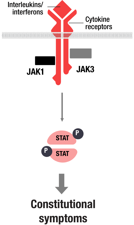
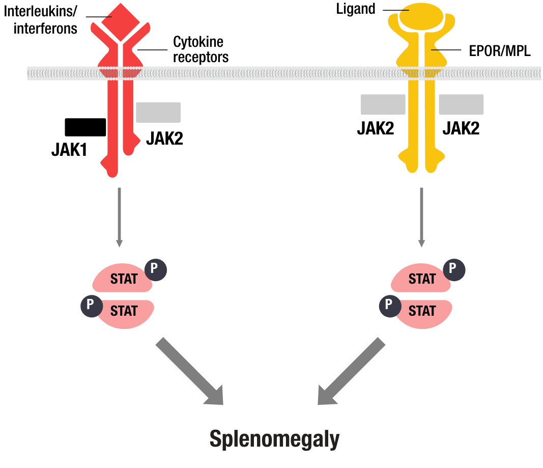
OVERACTIVE JAK-STAT PATHWAY SIGNALLING IS A KEY DRIVER OF MF1,2

- Constitutional symptoms driven by increased levels of circulating inflammatory cytokines are increasingly recognised as being attributed to overactive JAK1 signalling1

- Overactive JAK2 signalling has been implicated in splenomegaly in patients with myelofibrosis (MF)2
PATHWAYS BEYOND JAK-STAT ARE BEING EXPLORED TO FURTHER UNDERSTAND THE DRIVERS THAT MAY CONTRIBUTE TO KEY MANIFESTATIONS OF MF, INCLUDING CYTOPENIAS3,4
ACVR1/ALK2
The activin A receptor type 1 (ACVR1) gene, which encodes for the activin receptor-like kinase-2 (ALK2), is involved in bone morphogenetic protein (BMP) signalling. ALK2 activation leads to increased hepcidin production, which in turn promotes anaemia.5,6
BCL-2/BCL-XL
Myeloproliferative neoplasm (MPN) cells with Janus kinase 2 (JAK2) mutations are believed to be dependent on B-cell lymphoma 2 (Bcl-2) and B-cell lymphoma-extra large (Bcl-xL) proteins for survival. Overexpression of these proteins can help mutated cells avoid apoptosis and continue proliferating.7,8
BET
Bromodomain and extra-terminal domain (BET) proteins are epigenetic readers that are believed to contribute to the progression of MF due to their involvement in several signalling pathways. Activation of BET has been shown to increase nuclear factor kappa-light-chain-enhancer of activated B cell (NF-κB) signalling, thereby increasing inflammation.9
PI3K/AKT
Aberrant activation of the phosphatidylinositol 3-kinase (PI3K) and protein kinase B (Akt) pathway is present in cancer and other diseases involving immune deficiencies and tissue overgrowth. Akt activation leads to proliferation of normal or malignant cells while the activation of the PI3K pathway can promote the production of proinflammatory cytokines.10,11
TELOMERASE
Telomerase is an enzyme that maintains telomere length in rapidly dividing cells. Upregulated telomerase activity, which has been observed in myeloproliferative neoplasm (MPN) cells, is thought to enable telomere length maintenance and cell survival in rapidly dividing cells.12
TGF-β
Transforming growth factor beta (TGF-β) expression is elevated in primary myelofibrosis (PMF) patients compared to people without PMF. In addition to inducing fibrosis, TGF-β has been shown to inhibit haematopoiesis and negatively affect erythroid differentiation.13
BET, bromodomain and extra-terminal domain; Bcl-2, B-cell lymphoma 2; Bcl-xL, B-cell lymphoma-extra large; BMP, bone morphogenetic protein; EPOR, erythropoietin receptor; JAK, Janus-associated kinase; JAK-STAT, Janus kinase-signal transducer and activator of transcription; MF, myelofibrosis; MPL, myeloproliferative leukemia virus; MPN, myeloproliferative neoplasm; NF-kB, nuclear factor kappa-light-chain-enhancer of activated B cell; P, phosphorylation; PMF, primary myelofibrosis; STAT, signal transducer and activator of transcription; TGF-β, transforming growth factor beta
References: 1. Mascarenhas J. Leuk Lymphoma. 2015;56(9):2493-2497. 2. Tremblay D et al. Ann Hematol. 2020;99(7):1441-1451. 3. Schieber M. et al. Blood Cancer J. 2019;9(9):74. 4. Naymagon L, Mascarenhas J. HemaSphere. 2017;1(1):e1. 5. Aykul S et al. J Clin Invest. 2022;132(12):e153792. 6. Valer JA et al. Cells. 2019;8(11):1366. 7. Tognon R et al. J Hematol Oncol. 2012;5:2. 8. Petiti J et al. J Cell Mol Med. 2020;24(18):10978-10986. 9. Kleppe M et al. Cancer Cell. 2018;33(1):29-43.e7. 10. Bartalucci N et al. J Cell Mol Med. 2013;17(11):1385-1396. 11. Fruman DA et al. Cell. 2017;170(4):605-635. 12. Tefferi A et al. N Engl J Med. 2015;373(10):908-919. 13. Ceglia I et al. Exp Hematol. 2016;44(12):1138-1155.e4.


